
keyence_sop
Table of Contents
Keyence Microscope Training SOP
Last edited: Haley (7/28/22)
Training Checklist:
- Safety: do not leave lights on, do not crash objectives, wear gloves if replacing bulb
- Microscope Setup
- Changing objectives
- Tilting the head
- Changing lighting elements
- Moving/positioning the stage
- At the Computer
- Initialize the XY stage
- Software lighting and image quality
- Depth up
- Measure
- Stitching
- Post Imaging: replacing objective, turn off microscope light, replace original glass
Overview:
- This training provides an introduction to using and operating the Keyence microscope including:
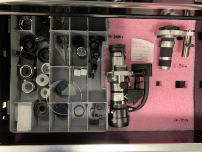
- Changing Objectives
- 0-50x and 20-200x
- 100-1000x
- Head tilt
- Stage movement
- Software
- Lighting
- Measure
- Depth up
- Image quality
- Optimize
- Stitching
- Safety
- General microscope use
- Maintenance
Safety
- Do not leave lighting elements on
- Do not crash the objectives
- If replacing the bulb in the MI-150 wear gloves to avoid getting oil on the halogen lamp bulb
Microscope Setup
At the microscope:
- Changing objectives
- Press the button on the top of the objective (right above the Y axis of rotation screw)
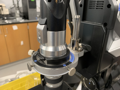
- Twist the camera body counterclockwise and carefully lift directly off. Set carefully on its side to avoid damaging the lenses in the camera assembly
- Loosen the large plastic wingnut screw located above the axis of rotation adjustment on the objective. (DO NOT ADJUST AXIS OF ROTATION SCREWS)
- Lift the objective straight up off the mounting post on the microscope body and place carefully in drawer located under Keyence computer.
- Remove the desired objective and lower over the mounting post on microscope body.
- Gently snug up the wing nut screw to clamp on the flat of the mounting post
- Gently lower the camera assembly onto the top of the objective
- Once lined up, rotate camera assembly clockwise until a sharp click is heard
- Make sure zoom knob is locked into a set zoom level detent
- Follow steps to initialize the XY stage
- Tilting the head
- The microscope head is able to tilt up to 90 degrees clockwise in order to better image samples. Caution should be taken to avoid crashing the objective into the sample or stage when head is tilted.
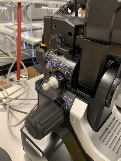
- Loosen the tilt lock knob a turn or two max (until the knob loosens)
- With a HAND ON THE MICROSCOPE HEAD rotate the locking lever counter clockwise to its vertical position
- Tilt the head to its desired angle using the graduations on the microscope body
- Lock the head in its tilted position using the large tilt lock knob. Make sure knob is snug before letting go of the head but do not overtighten
- If desired angle is greater then 60 degrees, pull the locking pin out away from the microscope before retightening the tilt lock knob. This should allow the microscope head to tilt up to 90 degrees. Be incredibly careful as the stage and objective will become very close at angles above 60 degrees.
- Changing lighting elements
- Switching to fiberoptic Epi-illumination for 100-1000x objective
- With the 100-1000x objective installed turn on the MI-150 fiberoptic lamp
- Install the end of the fiber into the lighting element of the objective. Light intensity is now controlled using the MI-150 instead of the microscope
- Switching the stage glass for backlighting
- Some clear or translucent samples may benefit from backlighting during imagery
- Remove the black and white disk in the center of the stage being careful not to bump the objective
- In the drawer containing the objectives there is also a class disk which can be placed in the stage center.
- Moving/positioning the stage
- X,Y movement
- X and Y positioning of the sample can be done using the joystick on the console
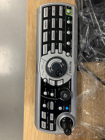
- Double clicking a location on the sample on the Keyence screen will center that part of the sample
- Z movement
- Unless the sample being imaged is physically large enough to necessitate moving the stage down it should be kept raised in its highest position with focusing being taken care of by raising and lowering the objective.
- To move the stage in Z, the silver locking ring with knurling must be rotated counterclockwise at most a rotation or two (until the adjacent knob on the left side of the microscope can rotate. Some tension from the locking ring is needed to keep the stage from lowering itself)
- The knob on the left side of the microscope body can then be used to raise or lower the stage
- Once the stage has been positioned, gently snug up the silver locking ring on the right side of the microscope to lock the position in place. DO NOT OVER TIGHTEN
- Focusing the lens
- The lens also moves in z and can be controlled by either the center dial on the control or the computer mouse wheel.


At the Computer:
- Initialize the XY stage
- The XY stage needs to be initialized every time a objective is switched or the microscope is power cycled
- The microscope should prompt the user after objective is switched but can be done manually through “settings” → “initialize XY Stage”
- Be sure the objective is locked in a zoom detent and the XY stage is raised and locked to its highest position
- Be sure the black side of the XY stage disk is facing up and is fully in place
- click “initialize XY Stage” and the microscope will initialize automatically by homing the stage
- Software lighting and Image Quality
- Within Software lighting two tabs are available:
- Brightness
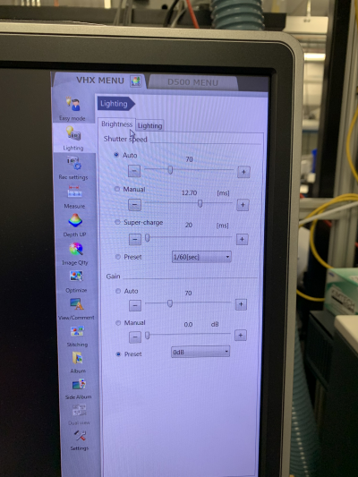
- Adjust shutter speed and camera settings (set to auto)
- Lighting
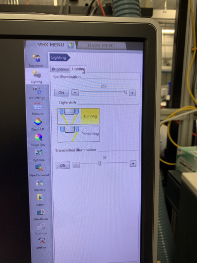
- Epi-illumination
- Adjust/turn on internal objective illumination (press “light” on console to toggle)
- Transmitted illumination
- Adjust/turn on sample backlighting (requires the use of glass stage ring)
- When 100-1000x objective is installed Epi-illumination is handled by the fiberoptic MI-150 located next to the microscope
- Increased contrast can be achieved by turning on HDR under the Image Quality tab
- Depth up
- The Keyence microscope is capable of taking hundreds of images as it raises the objective to capture an image larger depth of field. This is useful for observing objects that do not lie on a flat plane and would normally not be completely in focus. This image also contains depth mapping data which can be used to not only take XY and Z measurements, but also create a 3D mesh file of the scanned object.
- Select “depth up” from the right hand side bar then “quick composition and 3D”
- Focus the microscope slightly below the lowest point on the object
- Click “3D Display” on the Keyence control pad and the microscope will scan up the object and create a 3D file which can be analyzed for depth.
- Measure
- Measurements can be taken by selecting the “Measure” tab to measure the current view of the microscope or after a Depth up scan by selecting the “measure” tab after scan is complete
- Select the desired tool and desired points to measure from on the sample. The microscope will automatically display the length of lines or radius of arcs/circles drawn


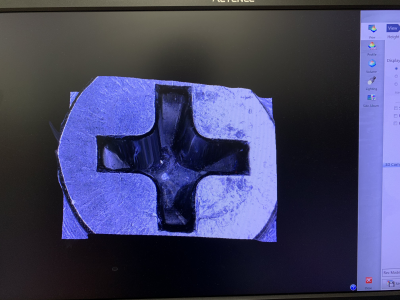
- Stitching
- Center surround stitching
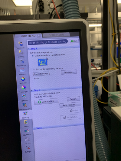
- Center the sample in the frame and select “stitching” from the sidebar
- select “stitch around the current position”
- click “Start stitching” and press enter to stop the stitching whenever the image collected reaches the desired size.
- Stitch after specifying the area
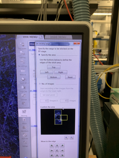
- Click select range
- Position the microscope as the minimum x position and select left, then maximum x position and select right
- Position the microscope as the minimum y position and select top, then maximum y position and select bottom
- Select start stitching and the microscope will automatically stitch together an image from the bounds set


Post Imaging
- If using another objective, please replace the 20-200x objective and initialize the microscope before leaving
- Be sure to turn off the microscope light as well as the fiberoptic MI-150 when imaging is finished
- If different light diffusers or polarizers are used during imaging please replace the original glass before leaving
Keyence microscope Quick Review
Tool Lead:
Contact: andrewfurst@ucsb.edu
Safety
- Do not leave lighting elements on
- Do not crash the objectives
- If replacing the bulb in the MI-150 wear gloves to avoid getting oil on the halogen lamp bulb
Safe Operation Procedures Review
keyence_sop.txt · Last modified: 2022/07/28 19:35 by haley